

Most GWAS loci stop being interesting the moment they are found. You get a region, a lead SNP, a nearest gene, and then twenty years of everyone citing the region as if it were the final answer. The CD40 locus at chromosome 20q13.12 has been like that. It has been associated with multiple autoimmune conditions including rheumatoid arthritis, Graves' disease, multiple sclerosis and inflammatory bowel disease for well over a decade, and CD40, encoding a costimulatory receptor of the TNF receptor superfamily carried on B cells, is so obvious a causal gene that nobody felt much pressure to go further.
A new preprint from Soumya Raychaudhuri's lab has now ripped apart this locus, plucking one base at a time, and arrives at an impressive list of insights. The authors pinpoint exact base that drives the genetic signal, its effect on the protein, leaving the transcription undisturbed, the cell type the effect lives in, and which cell state within that cell type. It's been a while a functional follow up of a common variant GWAS locus felt so satisfying to read.
The CITE-seq datasets and what the authors were trying to do
The starting point of the journey is CITE-seq, and the authors use it for something the technique was not built for. In CITE-seq you tag cells with antibodies that carry a short DNA barcode, then sequence the barcode along with the entire transcriptome, so every single cell comes back with its genome-wide gene expression and a count for each surface protein tagged on its surface. Almost everyone who used this technique has used the protein counts the way you use a flow cytometry gate, to say what kind of cell this is. The protein counts were designed to be mere labels.
The authors here however treated the counts as biological measurements. If you have the same panel across hundreds of genotyped individuals, the count for each protein on each cell type becomes a quantitative trait that you can correlate with genetics like any other trait. The authors pooled four CITE-seq datasets from three published studies comprising more than a million peripheral blood cells from 356 individuals and mapped genetic associations to 148 surface proteins across eight cell types.
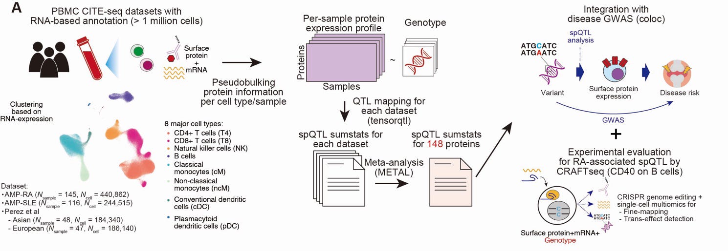
The reason for going after surface proteins is that they are what defines a cell state in the first place. A B cell announces what it is currently doing on its own surface, and other cells pick up that message and react. Most of the published protein QTL studies used serum or bulk tissue, and they inform nothing about the specific cell type the protein came from. CITE-seq offered a unique opportunity for the authors to study the genetic effects on proteins that sit on specific cell types.
What they found, and why they picked CD40
Their analysis revealed ~200 surface-protein QTLs (spQTLs) across 78 proteins, of which 19 proteins share genetic signals with an autoimmune GWAS locus with the same variant driving both the disease risk and the protein level change. Nineteen proteins is nineteen possible stories, and CD40 was one of them.
CD40 piqued the authors' interest as it stood out as the most clinically relevant protein. It's expressed on the B cell surface and it drives memory formation, proliferation and class switching. It binds to the CD40 ligand (CD40L) expressed on T cells, establishing the handshake between B and T cells that marks the activation of B cells and further differentiation. CD40 targeting is already in clinical trials.
The CD40 signal was among the strongest. The lead variant is rs1883832; each copy of its T allele tracks with roughly 20% less CD40 protein on B cells. The effect size is big for a common variant and the locus is the same one mapped in the rheumatoid arthritis GWAS, and also in Graves' disease, multiple sclerosis and inflammatory bowel disease. The interesting part of the finding here is the variant did nothing to the CD40 mRNA. A conventional eQTL study would have missed this signal.
Fine-mapping
Here the authors run into the wall that happens for any GWAS locus. An association locus represents a block of the genome where any of the multiple variants that are inherited together could be the disease culprit. This is not a GWAS-specific weakness. It applies to QTL studies as well. rs1883832 being the lead variant is a statement about the block, not the causal variant.
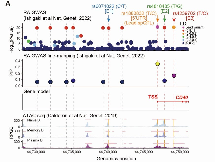
Statistical fine-mapping helps, but only to some extent. It narrows the block to a list of high confidence variants, but it doesn't break the linkage disequilibrium. Taking the fine-mapping results from their earlier GWAS of rheumatoid arthritis, the authors narrowed the list to ten candidate variants, all highly significant and none of them excluded.
The conventional next move is annotation. The authors overlaid the variants on the regulatory map of B cells, which annotates the places in the genome where chromatin is open. Three variants overlapped, all away from the lead variant. They added a fourth, the lead variant, as it lay inside the CD40 transcript, in the 5' UTR. So four candidates moved forward to the next step, three chosen because a regulatory logic nominated them and one because it sat within the transcript.
Base editing in Daudi to pinpoint the variant
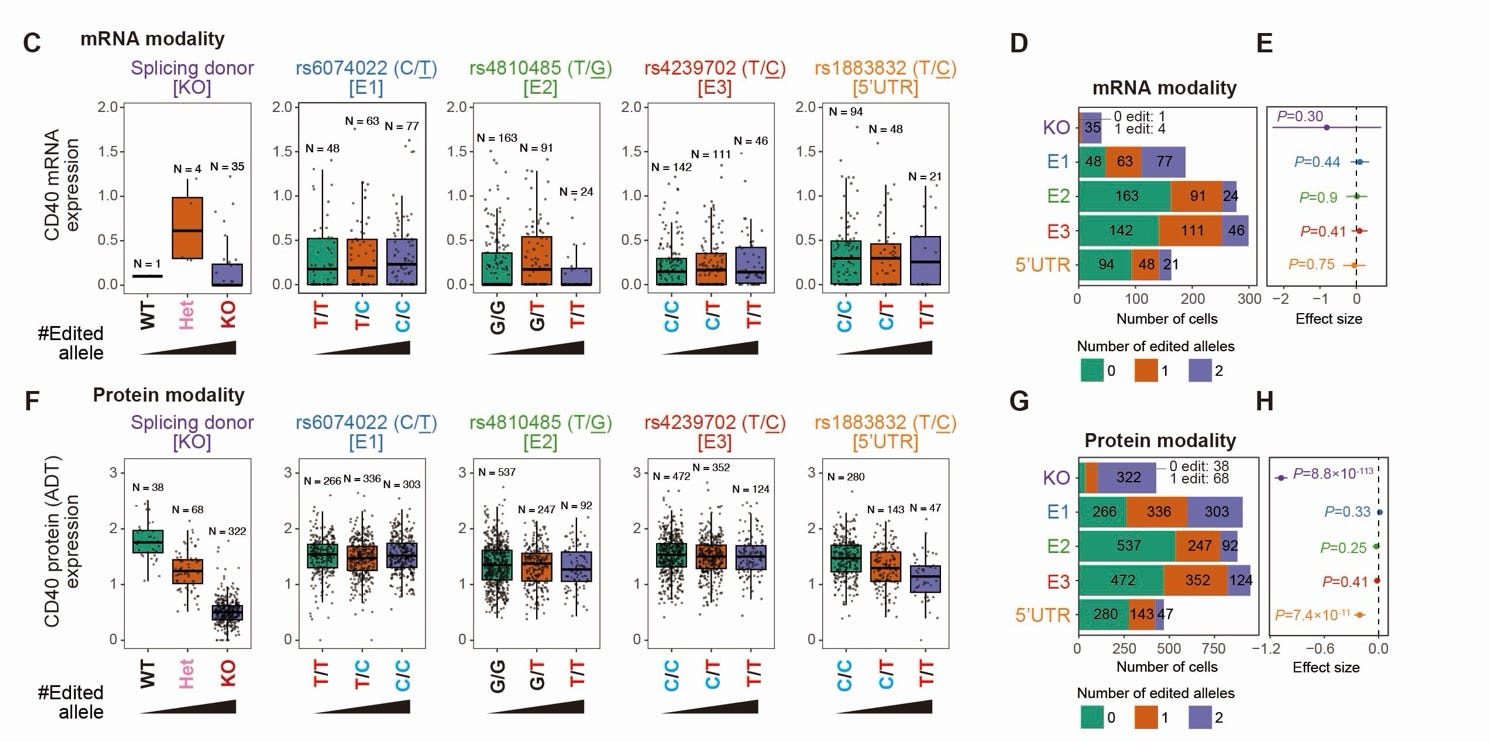
The authors used a brilliant approach to identify the causal variant. They disrupted one variant at a time while holding others constant and checked the outcome. Base editing helped accomplish this easily. In Daudi, an isogenic Burkitt lymphoma cell line derived from B cells, the authors edited one base at a time to measure the changes in CD40 surface protein, along with a positive control where CD40 is fully knocked out. Their readout was CRAFTseq, a method this same group published earlier, which reads the DNA of the edited region, the whole transcriptome and the surface proteins from within each single cell. It helped to turn a challenge of gene editing to their advantage. Editing has a downside, which is it is never complete, and often you end up with a mixture of edited and unedited cells. When all measurements (DNA, RNA and protein) happen within each cell separately, this is an advantage. Within an assay, you get natural comparators of different genotypes offering dose-response curves.
What did they find? None of the four candidates moved the CD40 mRNA significantly. And on the protein, one candidate, the one sitting on the transcript, had a significant effect, reducing the protein by about a fifth per copy of the T allele. The usual suspects (open chromatin variants) did nothing to either mRNA or protein.
The mechanism through which the 5' UTR variant directly reduced protein levels is a beauty. The variant sits one base upstream of the start codon, right on the Kozak motif, the short sequence the ribosome uses to recognise where to begin translating. The ribosome loads at one end of the transcript and begins scanning. It doesn't always stop at the first AUG codon it bumps into. It's the Kozak sequence next to AUG that tells the ribosome to stop and begin. Weaken it, and a fraction of the ribosomes scan straight past, producing no protein at all. The transcript is there in normal numbers, it's just that fewer proteins get translated from it. That's why the variant is invisible to transcription but visible to translation.
The mechanism itself is not new. In 2005, Jacobson and colleagues had the same variant in hand, already associated with Graves' disease, and ran an experiment. They placed the DNA template in a tube, a cell-free transcription and translation system, and measured how much CD40 protein was produced. The T allele made 15.5% less than the C allele. They then looked at B cells from people carrying each genotype and saw the same effect scaling with dose, 13.3% less CD40 in CT and 39.4% less in TT compared to CC. They called it translational pathophysiology. What they could not do at that time was pinpoint that this variant is the causal one driving Graves' disease risk. The mechanism and the exact base inferred by Jacobson et al. turned out to be right, but nothing in their experiment told them whether this base, rather than the nine sitting beside it, drove the protein change.
Validations
With the causal variant caught, the authors further validated their results using independent experiments. A larger editing experiment on rs1883832 alone in Daudi reproduced the finding. Then they used human donors with homozygous genotypes for both alleles (2 CC and 1 TT). In their B cells cultured with CD40L, they flipped the allele to T in the CC donors and to C in the TT donor, and observed the effects. Same result: protein down in proportion to the number of T alleles and no effect on mRNA.
Characterizing the trans-effect
Now they had nailed the causal variant. Next they went looking for what else in the transcriptome moved with this variant. As the CRAFTseq dataset from the edited primary B cells already had surface protein expression and the whole transcriptome data, they looked for trans-effects of the CD40 variant. 213 genes and 68 surface proteins were associated. Reassuringly the genes were enriched for CD40 signalling pathways and interestingly, many were established RA GWAS risk genes.
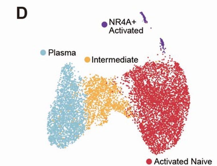
The dataset also let the authors cluster the cells to find subsets that represent different states of B cells. Looking at the trans-effects within each subset, only one subset showed the trans-effects. By contrast, all subsets showed the cis-effect. This suggests that the trans-effect is downstream of CD40 activation and that only a subset of the B cells are in that activated state. Their subsequent experiment supported exactly that.
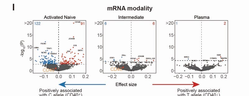
To test that directly, they had to take away CD40L, the T cell ligand that engages CD40 and is supplied in the culture. That is not possible in primary B cells, which don't survive without it, so the authors went back to Daudi. Comparing across the genotypes in Daudi with and without CD40L, the trans-effects showed up only in the presence of CD40L.
Cell-state mapping
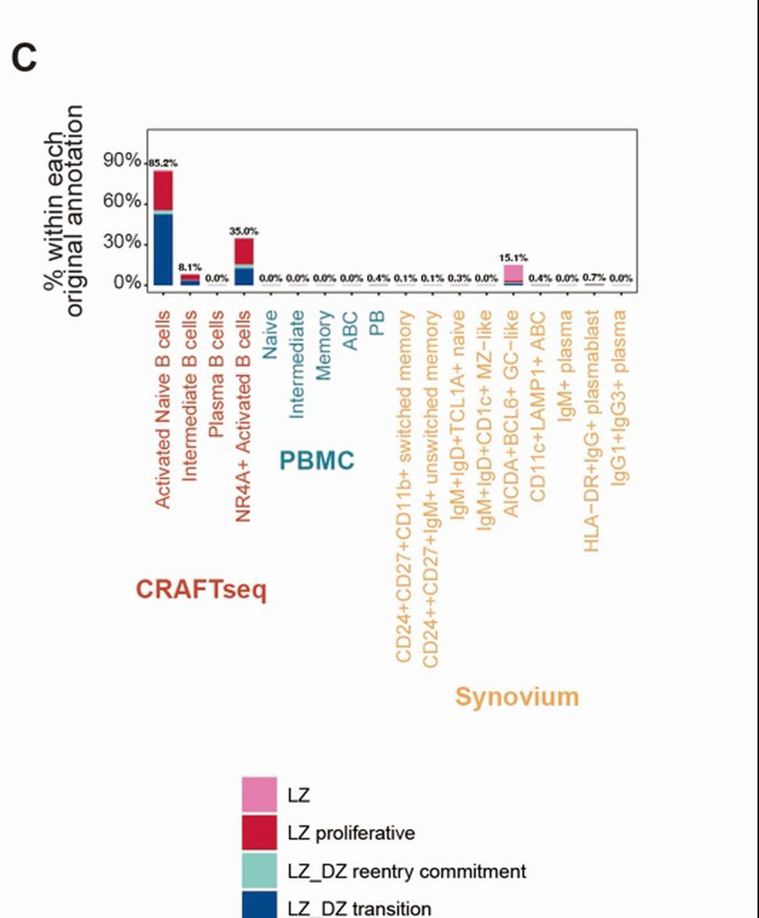
Having now established that only a specific subset of B cells manifests the trans effects, which are downstream of CD40-CD40L signaling, the authors went on to characterize the B cell states that map to this subset. The markers expressed by these cells told the authors that they resembled B cells enriched in the germinal centers, along with T cells holding the CD40L on their surface. The authors used a published cell type atlas based on healthy human tonsils, which contain germinal centers. As expected, >85% of the activated naive B cells mapped to light-zone states in the atlas. So, the CD40 variant is acting through signaling that happens only in B cells sitting in germinal centers, freshly activated through engagement with T cells via the CD40-CD40L handshake.
If so, the authors hypothesized that the trans effects cannot be detected in B cells in the peripheral blood, as such cells receive minimal CD40L stimulation compared to those in lymphoid organs. Running conventional trans-QTL mapping for the variant in B cells from population blood datasets, that's what they found: a solitary hit, with no relationship to the effects they had measured in the edited cells.
But what about the disease tissue? The authors next mapped B cells from inflamed RA synovial tissue onto the same atlas, and a subset of them — the ones annotated as germinal-centre-like — landed on the same light-zone states. The cells that carry the trans-effects are present where the disease happens.
Translational relevance
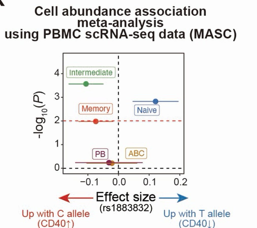
The authors conclude by asking one last question: the blood is blind to the trans effects, but what can it see? Perhaps the shift in the cell state itself, due to the CD40 reduction, that could be captured in the blood? Their intuition was right. Testing it in population blood data, they found that carriers of the T allele have fewer memory B cells and more naive ones — consistent with an older flow cytometry based GWAS that had linked this variant to IgD+ B cell counts. The finding makes sense, as CD40 is the ticket for B cells to graduate to memory cells. Lower CD40 protein would therefore shift B cell type proportions in the blood.
That leads to the translational insight. B cell depletion is already a tested idea to treat autoimmunity and it works, of course with caveats like infection risks. But the authors here raise an interesting question: do all B cell types need to be lowered? Maybe only the cell states that are relevant for the disease? The authors' work here supports that notion. In fact, treatments based on CD40 and CD40L targets are in trials. Iscalimab (CD40 blocker) works in Graves' disease, frexalimab (CD40L blocker) is in phase 3 for MS.1 But neither CD40 nor its ligand is a state-selective target. Every germinal center runs on CD40, including the one driving the vaccine response, and it is not confined to B cells. The authors' work opens the therapeutic path to find disease-selective B cell states to safely target to treat autoimmune diseases. Perhaps a CD40-like exercise is needed for a few other genes that were part of its trans network, and that would shed further light on the path.
Worth noting that the genetics and the drug pipeline point in opposite directions here. The T allele, which lowers CD40 protein, protects against RA and Graves' disease but increases the risk of MS, yet frexalimab, which blocks the same axis, is the most advanced compound in the class and it is in phase 3 for MS. One reconciliation is that a variant acts across a lifetime on who develops a disease, while a drug acts on disease already established, and a target can run in different directions on those two questions.
Thanks for the clean explanation